Click on the different blocks to read the full project description.
Imaging and Tracking Radiolabeled Stem Cells for Targeting Glioblastoma in Mouse Brain Models
We will perform in vivo SPECT Imaging of Stem Cells, functionalized with radiolabeled mesoporous silica nanoparticles for dynamic tracking of the most aggressive primary malignant brain tumor: the glioblastoma multiforme. The study will take advantage of the excellent performaces provided by the state-of-art MRC-SPECT-II system, developed in our lab.
[3-D rendering of fused SPECT/CT images of a mouse head acquired with MR-SPECT-I and Inveon microPET/SPECT/CT. Two small populations of radiolabeled stem cells injected in two hemisferes are visible in the image]
Statistical Techniques for the Design and Optimization of Complex Imaging Systems
Given the increasing complexity of modern radiological imaging systems, how do we know whether a given system has the most efficient configuration for collecting useful imaging information? and if not, how do we optimize the system that is characterized by hundreds or thousands of system parameters and by an infinite number of possible combinations? We have answered developing a statistical technique, based on the Modified Uniform Cramer-Rao bound (M-UCRB), to model the responses of any radiological imaging system to an arbitrarily given object, and to predict the properties provided by the system. Then we have integrated it in a general system optimization strategy that could accommodate almost any system parameter. It implements a systematic search through a very large parameter space to construct designs that provide the most statistical information possible and have low variances.
[ The Inverted Compound Eye Gamma Camera design as a challenging platform for applying the hardware optimization technique]
Hybrid X-ray Fluorescence, Luminescence and Transmission Computed Tomography
We develop a hybrid imaging modality that relies on X-ray fluorescence (XF), X-ray luminescence (XL), and X-ray transmission (XCT) measurements to produce 3-D multicolor images for guiding and monitoring nanoparticle-mediated microbeam therapy. During the microbeam therapeutic delivery process, a highly collimated X-ray beam irradiates the object. The pre-administrated metal-containing nanoparticles (NPs) preferably absorb the X-rays to produce therapeutic effects or directly from radiation effects induced by low-energy secondary electrons. Combining these three imaging techniques would provide a unique tool for guiding therapeutic delivery with highly detailed spatial and functional information: (1) the XF signal could determine the distribution of the NPs in the object, (2) the XL signal could provide quantitative information and indirect spatial mapping of the scintillation process induced by the X-ray irradiation of nanophosphors conjugated with photosensitizers and,finally, (3) the micro-CT images provide structural details of the object for confirmation of the delivery of the beam to the target area.
[ Preliminary imaging studies of a NP-loaded double-tube phantom that was filled with yttrium oxide nanoparticles in powder on the inside and sodium bromide aqueous solution on the outside]
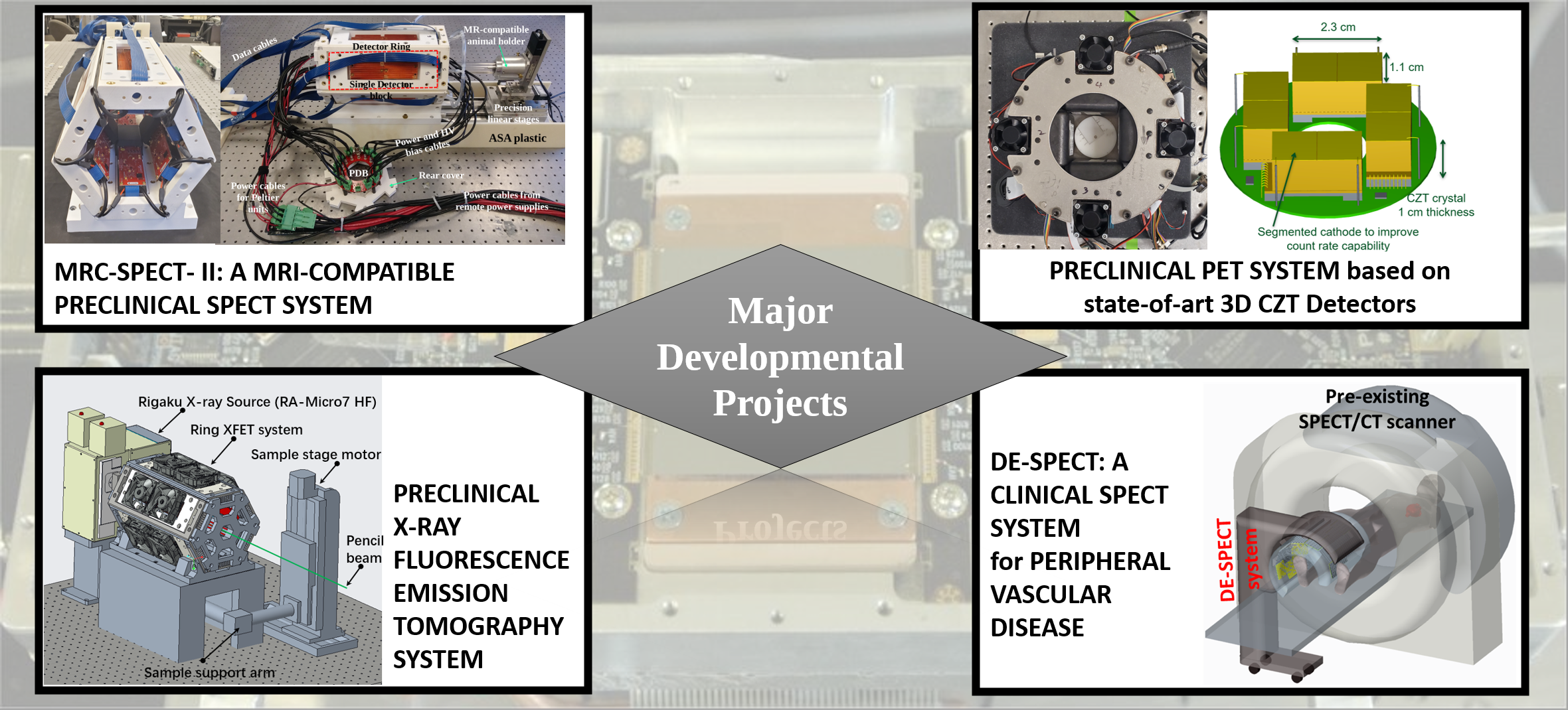
A Prototype Ultrahigh Resolution PET Imaging System based on state-of-art 3D-sensitive CZT Detectors
The preclinical PET system prototype employs a full ring of large volume 3-D position sensitive CZT detectors to offer an ultrahigh spatial resolution for small animals. Compared to the scintillation detectors employed in most of PET systems, the large volume CZT detectors used in this prototype offer an intrinsic spatial resolution of 0.5 mm FWHM in all three dimensions, along with an energy resolution of 1% FWHM at 511 keV. The detector has the capability of detecting multiple interactions induced by a single incident gamma ray with sub-0.5 mm resolution and an ultrahigh energy resolution (3 keV @ 200 keV, 4.5 keV @450 keV) for each detected interaction. This would allow the study of Compton kinematics to provide extra information on the dimensionality of the incident gamma rays.
[PD/AD mouse brain phantom: comparison between microPET (first row) and high-resolution PET (second row) reconstruction images]
Ultrahigh-Performance Gamma and X- Ray Imaging Detectors
We develop different high performance gamma ray imaging detectors for radiological imaging applications. We have combined compact CZT, CdTe and novel semiconductor material with custom-designed CMOS readout ASICs. These detectors provide excellent spatial, energy and timing resolution, and an adequate stopping power for medical applications.
MRC-SPECT-1 System
The MRC-SPECT-1 system is a preclinical MR-compatible SPECT system, fully customized for operation inside MR scanners as well as stand-alone platform. The system consists of ten CdTe imaging detector modules assembled in a compact ring with a distance of 156 mm between the surfaces of two opposite detectors. Each detector has four CdTe detector hybrids having each one 64 × 32 pixels of 350 µm × 350 µm pitch size for an overall dimension of 22.5× 11.2 × 2 mm^3 with four bump-bonded 2048-channel readout ASICs. Each detector is coupled with four 300- or 500-µm diameter pinholes. The SPECT detector ring is installed in a one-piece non-metal gantry with an outer diameter of 25 cm.
Imaging and tracking T-cells for brain cancer immunotherapy
A collaboration with Dr. Ed Roy's group at the University of Illinois
[3-D rendering of a fused SPECT/CT image of a mouse head. A small number (down to 1500) of radiolabeled T-cells are visible in the image]
Imaging of pancreatic beta-cell with combined SPECT/MR systems
A collaboration with Dr. Chin-Tu Chen's group at the Department of Radiology of the University of Chicago
3-D mapping of naturally occurring trace-metal in the brain
A collaboration with Dr. Patrick La Riviere's group at the University of Chicago
[3-D rendering of CT images from a Zebrafish Sample with 1% osmium tetroxide acquired using our X-Ray Stimulated Emission Tomography System]
Superhigh resolution PET imaging of mouse brain
A collaboration with Dr. Y-C. Tai's group at the Washington University in St Louis, and Dr. Q Z Li's group at the Massachussetts General Hospital