EQUIPMENT & SETUPS
Functional X-ray Imaging Benchwork
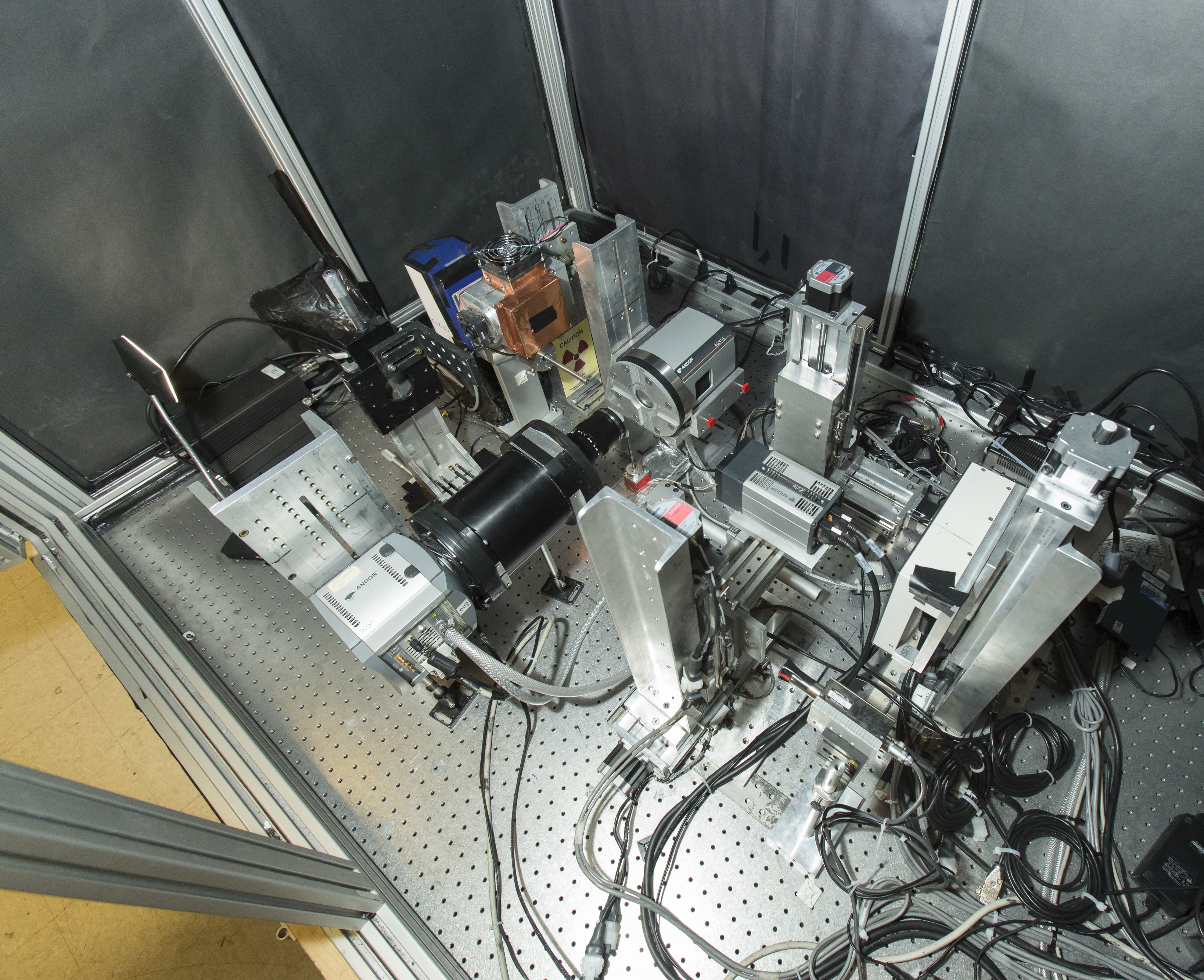
The X-ray fluorescence (XF), X-ray luminescence (XL), and X-ray transmission (XCT) imaging benchwork consists of:
(1) a monochromatic source that delivers a pencil beam of 17.4 keV X-rays with diameter tunable from 30 to 100 µm,
(2) a conventional micro-focus (25 µm) polychromatic X-ray source equipped with multiple filter configurations,
(3) a direct-detection deep-depleted X-ray CCD camera coupled to user-adjustable (slit or multiple-pinhole) apertures,
(4) an iXon Andor EMCCD camera with various optical filters, and (5) a Zyla 5.5 CMOS X-ray camera with a pixel size of 6.5µm.
The DE-SPECT SYSTEM
The Dynamic Extremities (DE-) SPECT system is a stationary and organ-dedicated clinical SPECT system based on 48 1-cm thick 3D-position-sensitive Cadmium Zinc Telluride (CZT) detectors and dynamic dual-field-of-view synthetic compound-eye (SCE) collimators. The scanner is designed and optimized to perform hyperspectral imaging of lower extremities in patients suffering of peripheral vascular diseases (PVD).
The MRC-SPECT-2 SYSTEM
The MRC-SPECT-2 system is a MR-compatible preclinical SPECT imaging system. The system presents 24 2-mm thick CZT crystals and a compound-eye collimator with 96 knife-edge pinholes. More details about the multi-detector readout circuitry can be found HERE. The system is currently installed at the Integrated Small Animal Imaging Research Resource (ISAIRR) at University of Chicago.
The ALPHA-SPECT-MINI SYSTEM
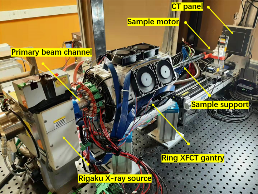
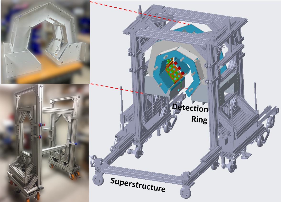
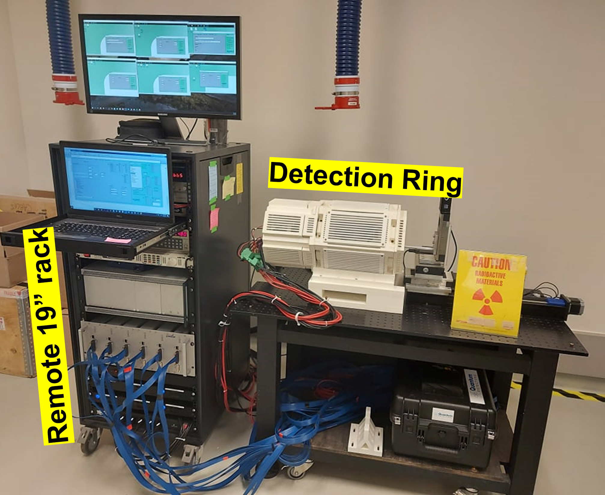
The Alpha-SPECTmini system is a high-performance small-animal system for XFCT and targeted alpha therapy imaging applications. The system is based on 24 1-mm CdTe detectors and it is equipped with interchangeable multi-slit and high-energy multi-pinhole collimators. The system is currently installed at the Department of Radiology and Radiological Science at the Johns Hopkins Medical School.
X-RAY SOURCES
1) Oxford Instrument, ultrabright microfocus X-ray source with Mo anode, capable of continuously operating at 90 kVp at 60 W with a focal spot size < 50 µm
2) TWO Apogee Oxford Instrument, polychromatic microfocus X-ray sources with W anode, capable of continuously operating at 50 kVp at 1 mA with a focal spot size of 25 µm
3) Xenocs GeniX 3D , a monochromatic X-ray source with Mo anode that delivers a pencil beam of 17.4 keV X-rays with a tunable diameter from 30 to 100 µm
4) XOS X-beam, a monochromatic X-ray source with Mo anode that delivers a pencil beam of 17.4 keV X-rays with a tunable diameter from 30 to 100 µm
5) Hamamatsu Microfocus L8121-03, a polychromatic microfocus X-ray source with W target, running up to 150kV, 10 W.
LEICA DMS300
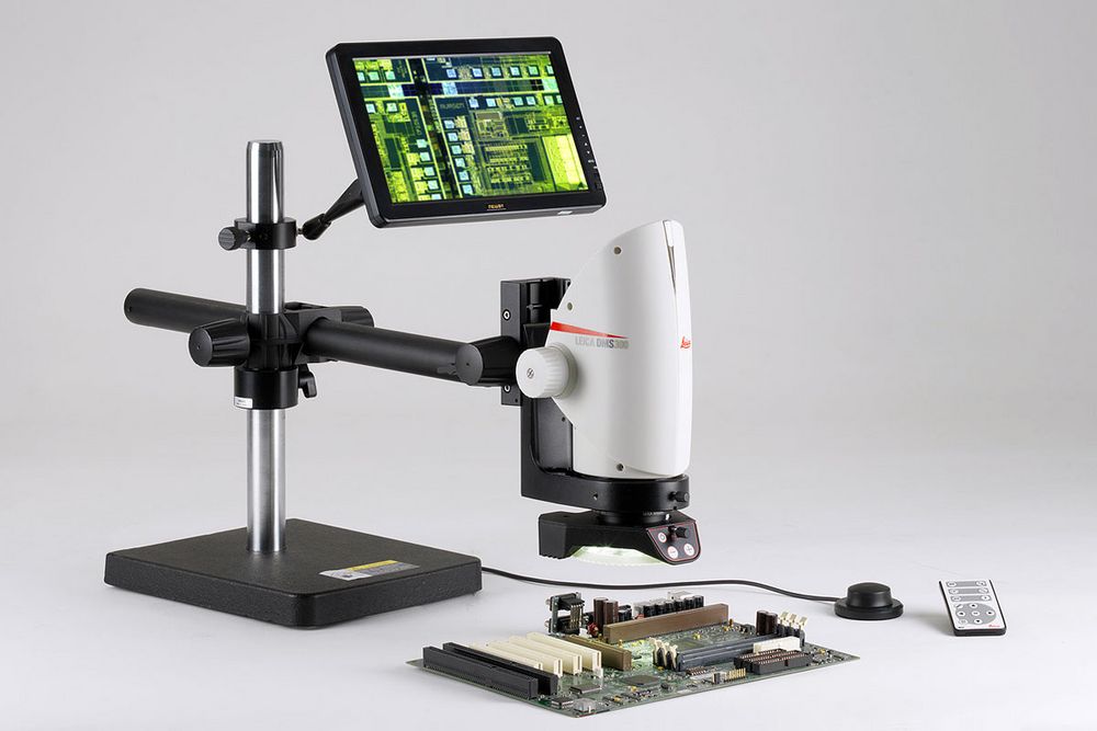
Digital microscope system with integrated high-end optics and high-performance digital camera, complete with LED ringlight and swing-arm system. Optimized for digital inspection and documentation.
GAMDIAS TELOS P1
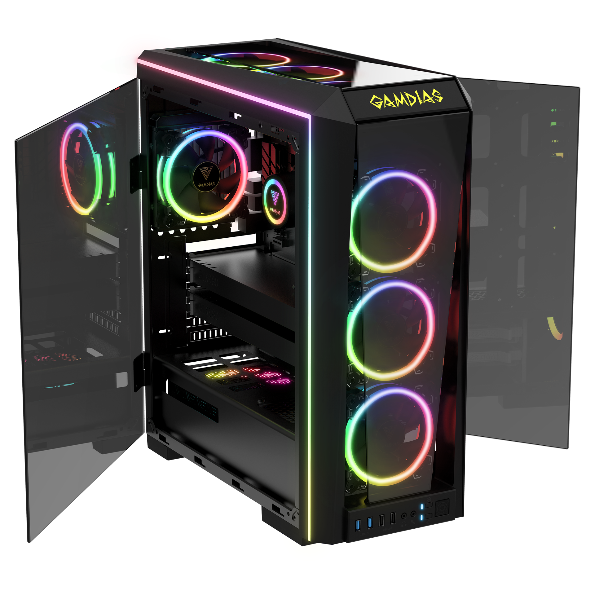
Two Gamdias Telos P1 servers equipped with the RTX3090 GPU for GPU-accelerated computing applications.
MAKERBOT REPLICATOR Z18
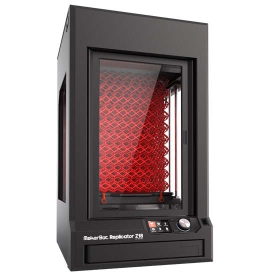
The Makerbot Replicator Z18 is an extra-large, professional 3D printer for ultra-tall concept models and prototypes.